
Eye health matters a lot in daily life. Many people face issues like cataracts or myopia that need precise checks. DGHA stands out as a handy device that helps doctors measure eyes accurately without much hassle. This post covers what DGHA is, its workings, features, benefits, and more. Readers will find straightforward info on this tool, including its role in eye care. Expect simple explanations, tips on usage, and comparisons to similar devices. By the end, clarity on DGHA’s place in modern ophthalmology will emerge.
DGHA, often called the Scanmate A, comes from DGH Technology. It serves as an A-Scan ultrasound gadget for eye pros. Compact and easy to carry, it connects to computers via USB. Doctors use it to gauge eye parts like axial length, anterior chamber depth, and lens thickness. Such measurements aid in planning surgeries or tracking conditions. The device blends tech with user-friendly design, making it popular in clinics worldwide. Its accuracy helps avoid errors in treatments.
What is DGHA?
This section gives a basic overview of DGHA, explaining its core purpose and design.
DGHA refers to the DGH 6000 Scanmate A, a specialized ultrasound instrument in ophthalmology. Made by DGH Technology, Inc it focuses on A-Scan biometry. The tool looks like a small box with a probe, weighing under a pound. It needs a Windows computer to run its software. Eye specialists pick it for quick, reliable scans. Unlike bulky machines, DGHA fits in bags for mobile use. Its main job involves sending sound waves into eyes to capture data.
The company behind DGHA started in 1982. DGH Technology grew from a focus on ultrasound pachymeters to full A-Scan and B-Scan lines. Based in Exton, Pennsylvania, it aims at affordable, quality gear for eye care. Over years, innovations like portable designs set it apart. DGHA represents that progress, offering features once limited to big setups. Pros appreciate its balance of cost and performance in routine tasks.

History of DGH Technology Inc.
Here, a short look at the company’s background shows how DGHA came about.
DGH Technology Inc. began operations in 1982. Founder Earl Henderson saw needs in eye ultrasound tech. Early products centered on pachymeters to measure corneal thickness. Success led to expansion into A-Scans by the 1990s. The firm earned a name for durable, user-focused items. Milestones include portable models that changed clinic workflows. Today, it remains independent, serving global markets with consistent quality.
Growth came through listening to doctors’ feedback. In the 2000s, DGH added software improvements for better data handling. The Scanmate series, including DGHA, launched around 2010. It addressed calls for lighter, versatile tools. Federal approvals, like FDA clearances, boosted trust. The company’s story highlights steady innovation without rushing trends.
How Does DGHA Work?
This part breaks down the device’s operation in simple steps.
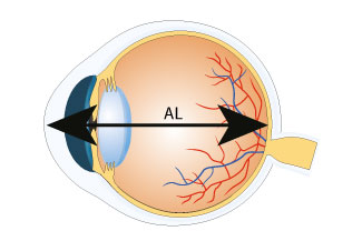
DGHA uses ultrasound principles for eye measurements. The probe emits high-frequency sound waves at 10 MHz. These waves travel through eye tissues and bounce back as echoes. The device times how long echoes take to return. Software then calculates distances based on sound speed in eye parts. For example, axial length gets figured from cornea to retina. Modes include contact, where the probe touches the cornea, or immersion, using a water shell to avoid pressure.
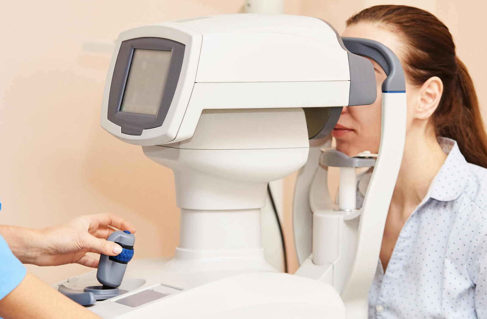
In practice, a tech attaches DGHA to a computer. Patient data goes in first. The probe aligns with the eye, guided by sounds and screen feedback. Good alignment earns stars – three means best. If pressure squishes the cornea, a lockout stops bad readings. After scans, IOL formulas compute lens powers for surgery. Reports print or save easily.
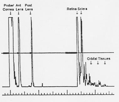
A-Scan biometry relies on precise echo detection. DGHA’s algorithm spots patterns for accuracy. It handles different eye types, like those with cataracts or implants. Immersion mode suits sensitive cases, reducing compression risks. Contact mode works faster for routine checks. The process takes seconds per eye, minimizing patient discomfort. Results store in databases for later review.
Key Features of DGHA
A quick note on standout elements that make DGHA useful.
One main feature involves real-time feedback. During scans, tones signal alignment quality. Stars rank each attempt, pushing for better ones. Compression lockout guards against squished corneas, adjustable for needs. The software packs IOL formulas like SRK/T or Haigis. Post-refractive options help after LASIK. Custom velocities allow tweaks for unique eyes.
Portability shines with USB power and light weight. It fits small spaces, ideal for travel clinics. Myopia reports track axial growth over time. Video saves let doctors replay scans. EMR exports keep records smooth. The probe, fixed at 10 MHz, ensures consistent output. Ranges cover 15-40 mm axial lengths, fitting most patients.
DGHA includes accessories like the Prager Shell for immersion. Software runs on standard Windows setups. Backups protect data. Reports customize with logos or notes. These traits combine for efficient, reliable work.
Benefits of Using DGHA
This section highlights advantages for users and patients.
Accuracy stands as a top benefit. DGHA’s pattern recognition yields repeatable results, with 0.03 mm precision in immersion. This cuts surgery surprises. Portability lets doctors scan anywhere, from offices to field sites. No need for heavy gear means lower costs. Patients enjoy quick, painless tests – no drops or long waits.
Software eases workflows. Multiple formulas in one spot speed planning. Progression reports aid myopia control in kids. Compatibility with EMRs reduces paperwork. The device’s affordability appeals to small practices. Training takes little time, thanks to intuitive guides. Overall, DGHA boosts efficiency without sacrificing quality.
Compared to older models, DGHA offers modern perks like video storage. It handles dense cataracts well, where optics fail. Patients with implants get accurate reads too. The tool’s durability means long service life. Pros report fewer errors, leading to better outcomes.
Applications in Eye Care
Brief info on where DGHA fits in daily practice.
Cataract surgery prep tops the list. DGHA measures axial length for IOL power picks. Accurate biometry leads to sharp vision post-op. In myopia management, it tracks eye growth. Regular scans spot changes early, guiding treatments like orthokeratology. Dense cataracts block light-based tools, so ultrasound steps in.
Post-refractive cases use special formulas. DGHA calculates for eyes after laser work. Diagnostic scans check anterior chamber depth or lens thickness. This helps spot issues like glaucoma risks. Pediatric care benefits from quick, non-invasive methods. Clinics use it for baseline data in routine exams.
Research settings employ for studies on eye dimensions. Its data exports aid analysis. In low-resource areas, portability brings advanced care. Overall, it supports varied needs in ophthalmology.
How to Use DGHA Step by Step
A short guide on operating the device.
Start by setting up. Connect DGHA to a USB port on a compatible computer. Install software from DGH if needed. Enter patient info like name and eye type. Choose mode – contact or immersion. For contact, apply gel to probe tip. Align probe perpendicular to cornea, listening for tones.
Watch the screen for waveforms. Adjust until three stars show. Capture multiple scans for averages. Software flags bad ones. Switch eyes if needed. Run IOL calculations by selecting formulas. Review results, then generate reports. Save or export data. Clean probe after use with approved wipes.
Tips include practicing alignment for speed. Use immersion for picky cases. Check batteries in portable setups. Software updates add features, so stay current. Training videos from DGH help beginners.
Comparison with Other A-Scan Devices
Here, a look at how DGHA stacks up against rivals.
Many A-Scans exist, like those from Sonomed or Quantel. DGHA excels in portability – lighter than desk models. Its USB setup beats standalone units for data ease. Accuracy matches gold standards, but at lower price. Some competitors offer optical biometry, yet ultrasound wins in opaque media.
Versus optical tools like IOLMaster, DGHA costs less and handles cataracts better. Optical skips contact, but fails in dense lenses software rivals in formulas, though some have more automation. Build quality holds up, with warranties backing it. Users note easier learning curve with DGHA.
In modular systems like Scanmate Flex, integrates for added B-Scan or UBM. Standalone rivals lack that flexibility. Repeatability studies show similar stats. Choice depends on clinic size suits mobile or budget setups.
Maintenance and Care for DGHA
Brief tips on keeping the device in top shape.
Regular cleaning keeps DGHA reliable. Wipe probe with soft cloth and disinfectant after sessions. Avoid harsh chemicals. Store in case to protect from dust. Check cables for wear monthly. Software backups prevent data loss. Calibrate as per manual, usually yearly.
DGH offers support via phone or email. Warranties cover defects for one year. Extend coverage for peace. Update software for bug fixes. Train staff on proper handling to avoid drops. In humid spots, use dehumidifiers. These steps extend life and ensure accurate reads.
Troubleshooting common issues: If no signal, check connections. Waveform noise might mean dirty probe. Software glitches? Restart computer. DGH manuals detail fixes. Proactive care minimizes downtime.
Future Trends in A-Scan Technology
This part discusses upcoming developments related to DGHA-like tools.
Advances point to AI integration. Future A-Scans might auto-analyze waveforms for faster results. Wireless connections could replace USB for mobility. Smaller probes suit pediatric use. Hybrid devices blending ultrasound and optics gain traction. DGH might add such features in updates.
Myopia control drives demand for tracking tools reports could expand with predictive models. Telemedicine links allow remote data shares. Sustainability focuses on eco-friendly materials. Regulations push for better data security. Overall, tech like evolves to meet growing eye care needs.
Research explores 3D mapping with A-Scans. Portable power sources extend field use. Cost drops make tools accessible globally. DGH’s history suggests continued improvements. Eye pros anticipate smarter, easier devices ahead.
Conclusion
In summary, DGHA proves a solid choice for eye measurements. Its mix of accuracy, portability, and ease suits many practices. From cataract prep to myopia tracking, it delivers value. Eye care keeps advancing, and tools like this play a key role. Pros seeking reliable gear find DGHA worthwhile. Patients benefit from precise, comfortable tests. As tech moves forward, expect more from such innovations.